I just had a necropsy done on an adult female pancake and thought I would share the results.
History of not eating for 2 days. Otherwise, active, drinking and producing normal stool. Seen alive and walking at 8am, found deceased at 12pm the same day. Body was immediately frozen as it would be 2 weeks before vet was back in town to be able to do the necropsy.
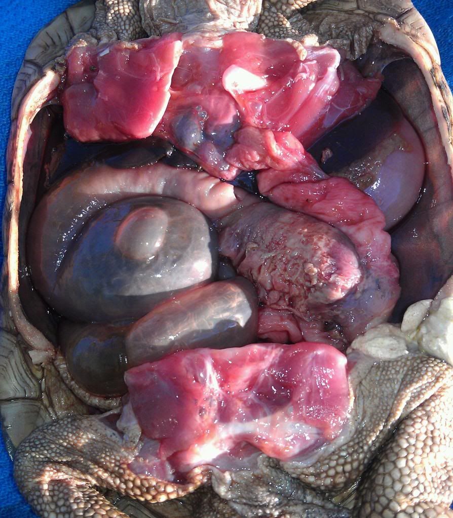
Necropsy results showed a 2 cm section of colon discolored and thickened. Once the colon was turned over, the underside showed very friable tissue with a small perforation. The inside of this section of colon was very dark in color and showed many fibrous layers that were easily peeled away. There was no evidence of a recent foreign body in or around the colon. There was also a large amount of bacteria filled free fluid in the entire body cavity.
What that means is, she had probably ingested something up to or over a year ago that damaged her colon. Whatever this was, she was able to pass but her body had to repair the damage. In doing this it stayed inflamed and kept adding layers of tissue. Eventually= (2 weeks ago) a perforation developed in the colon and she began leaking feces into the body cavity. This was full of bacteria and she became septic. This is what killed her.
He said he would only expect her to have lived maybe 2 days once she was septic. Had she not perforated the colon she would have eventually become obstructed and had the same end result. He said that had this problem been found before she was septic or obstructed, surgery would have been the only option. To resect that much of the distal colon would have been difficult and the odds would have been against her being able to recover at all.
I hate that I no longer have this beautiful girl. I'm grateful to know that I didn't miss something and that more than likely, I could not have saved her even if we found this and did the surgery. Because of her, I now have a greater knowledge of the anatomy and inner workings of tortoises and hopefully after seeing this, you do too.
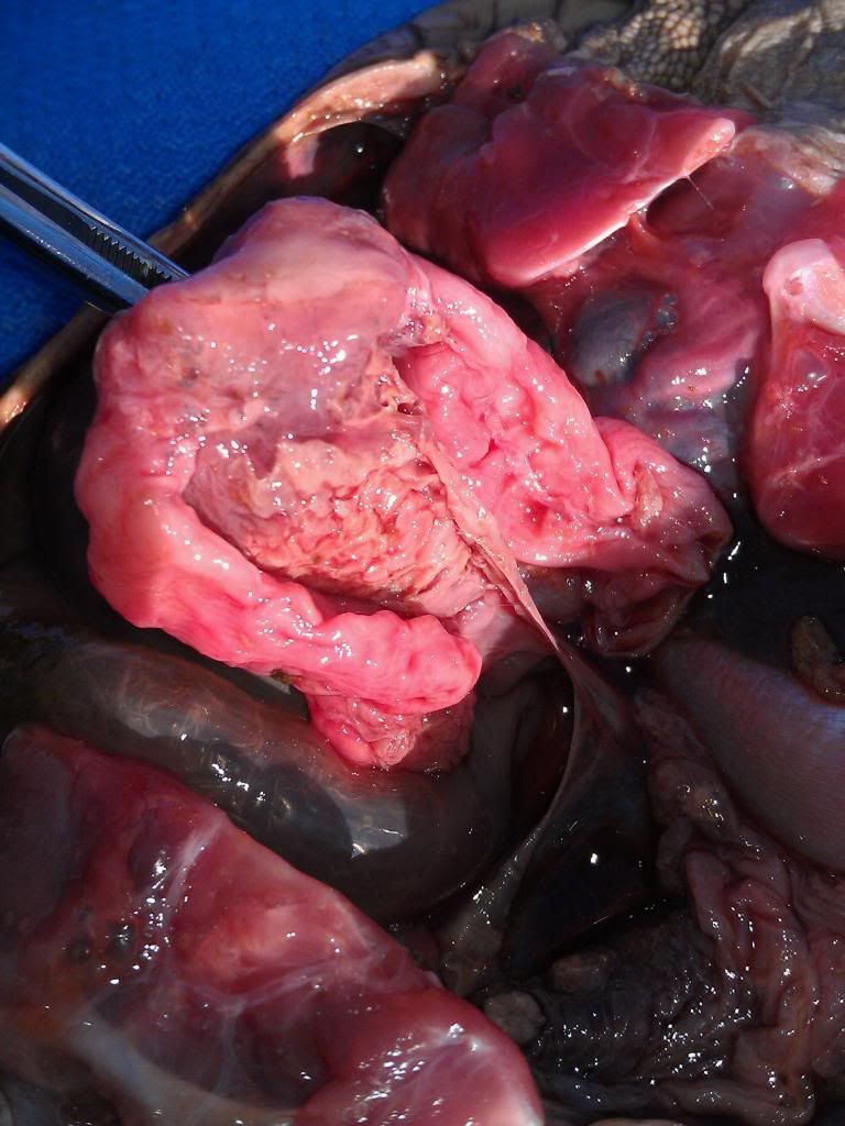
Large, thickened area of the top side of the colon in center of photo. This is after removing the fluid, fat and cutting colon free of the body attachments. Note, bubble is from gas building up in the secum post-mortum.
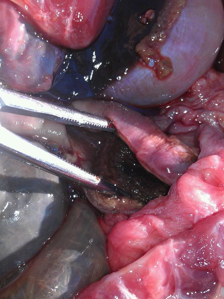
Underside of colon. Friable tissue where the perforation is.
Thickened and discolored area inside of the colon.
History of not eating for 2 days. Otherwise, active, drinking and producing normal stool. Seen alive and walking at 8am, found deceased at 12pm the same day. Body was immediately frozen as it would be 2 weeks before vet was back in town to be able to do the necropsy.
Necropsy results showed a 2 cm section of colon discolored and thickened. Once the colon was turned over, the underside showed very friable tissue with a small perforation. The inside of this section of colon was very dark in color and showed many fibrous layers that were easily peeled away. There was no evidence of a recent foreign body in or around the colon. There was also a large amount of bacteria filled free fluid in the entire body cavity.
What that means is, she had probably ingested something up to or over a year ago that damaged her colon. Whatever this was, she was able to pass but her body had to repair the damage. In doing this it stayed inflamed and kept adding layers of tissue. Eventually= (2 weeks ago) a perforation developed in the colon and she began leaking feces into the body cavity. This was full of bacteria and she became septic. This is what killed her.
He said he would only expect her to have lived maybe 2 days once she was septic. Had she not perforated the colon she would have eventually become obstructed and had the same end result. He said that had this problem been found before she was septic or obstructed, surgery would have been the only option. To resect that much of the distal colon would have been difficult and the odds would have been against her being able to recover at all.
I hate that I no longer have this beautiful girl. I'm grateful to know that I didn't miss something and that more than likely, I could not have saved her even if we found this and did the surgery. Because of her, I now have a greater knowledge of the anatomy and inner workings of tortoises and hopefully after seeing this, you do too.
Large, thickened area of the top side of the colon in center of photo. This is after removing the fluid, fat and cutting colon free of the body attachments. Note, bubble is from gas building up in the secum post-mortum.
Underside of colon. Friable tissue where the perforation is.
Thickened and discolored area inside of the colon.
